Role of Radiotherapy in Gynecological Cancer

Introduction:
Radiotherapy is the art of using ionizing radiation to destroy malignant tumors while minimizing damage to normal tissues ( J.coate).
CIN and Cancer Difference
Radiotherapy is the important modalities of therapy for human cancers apart from surgery. It is very useful in the treatment of many cancer specially female reproductive tract.

fibroscopic visualization technique for urinary bladder visualization

Mechanism action of radiotherapy

1)Radiation causes ionization of water of the cancer cells into hydrogen and hydroxyl ions which are toxic and causes damage to DNA of the cell and thereby cell death. [The target for radiation is injury to DNA, which is responsible for cell mitosis. So injury to DNA causes mitotic cell death.]
2)It stimulates fibrous tissue reaction in the surrounding stroma and resulting filorosis causes entanglement of the tumour cells and prevents local spread.
3)Reducing vascularity by radiation end arteritis.

Technique/Methods of radiotherapy

For gynecological cancers therapeutic
radiation is delivered as –
  • a) External radiation (Teletherapy)
  • b) Internal radiation (Brachytherapy)
a) Teletherapy- The goal of teletherapy is to ensure that radiation is delivered to the target tissue without affecting uninvolved tissues from distance (usually 100cm). Cobalt 60 is the common teletherapy source for external radiation; other one is cesium 137. External radiation is used to treat large volumes of tumours, lymph nodes, parametrium.
b) Brachy therapy (internal radiation)

Here source of therapeutic ionizing radiation is placed close to the treatment area, chief advantages of it is that a relatively high dose of radiation can be applied to a limited anatomic region.
Commonly used sources are caesium-137
Cobalt - 60
The application may be
  1. Intracavitary
  2. Interstitial Intracavitary – The devices for brachytherapy consists of hollow stem; intrauterine tandem, which is placed within the uterine cavity. Special devices used for vaginal placements are called vaginal ovoids or colpostats.
    Interstitial – Using needles, wires or seeds used as radio active sources within tissues.

    Indication of radiotherapy

    a) Cervical cancer- It is used in all stages. In squmous cell type, response is best for radiotherapy.
    b) Endometrial cancer- Adenocarcinoma response to radiohterapy.
    c) Vaginal cancer – Here radiotherapy is best than surgery.
    d) Vulval cancer – Generally radiothery is not used, as vulval skin is moist, Causing irritation & distort the shape of vulva.
    e) Orarian cancer – Actualy no role of radiotherapy but in dysgerminoma, responsed of radiotherapy is good.
    f) Choreocarcinoma – When metastasis to liver & brain.

    Uses of radiotherapy in specific cancer

    Cervical cancer: Treatment of cervical cancer is a prime example of successful application of radiotherapy.
    Radio therapy can be used in all stages of carcinoma of cervix but in stage 0 or pre-invasive carcinoma we do not use radiotherapy due to damage of ovarian tissue.
    In cancer cervix
     
    In early stage – option of stage 1 + upto stage II A
    Mode of treatment –
    a) Only surgery
    b) surgery + Radiotherapy
    c) Only Radiotherap
    But stage IIB & on wards option of mode of treatment
    •Only radiothery
    •Here surgery is not possible.
    Goal of external radiation – in cervical cancer is to Sterilize metastatic disease to pelvic lymphnode and parametrium and to decrease the size for cervix to allow placement of intracavitary radio active sources.
    It is also used as palliative radiation for
    advanced cervical cancer for-
    •Control of bleeding
    •Pain relief.
    Radiotherapy combined with chemotherapy specially platinum based cisplatin or carboplatin provides better survival.
    Dose of radiotherapy
    Bnachytherapy – in the form of intracavitary.
    The goal is to deliver a dose of 70-80 Gy at point A, which is situated 2 cm above the mucosa of lateral vaginal fornices & 2 cm lateral to the central uterine canal.

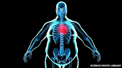
Obesity can decrease Vitamin D of your body

Over weight

People who have scored B.M.I (Body Mass Index) 30 or more than 30 are counted as obese person in medical science. This obesity or fattiness can decrease your level of vitamin D in your body.

Recently scientists have found that by researching  genetic data of human body and examine about 42 thousand people. They said that about 10% increasing of B.M.I (Body Mass Index) can cause decreasing of 4% vitamin D of total level.

fast food habit

It is considered that maximum amount of vitamin D is stored in fatty tissue of body. But in case of obese or fatty people have got more fatty tissue than normal. So, it caused obstacles in passing vitamin D trough fatty tissue to blood circulation in proper manner.  In relation with that condition Professor David Has-lam, Member of National Obesity Forum said, “ Food habit and genetic factors both are responsible for obesity of human body.”


In blood the normal level of Vitamin D is 50 nano mol per liter. If it decease less than 30 it will cause weakness of bone, Rickets of children and another bone diseases.

Human Body

                                                                                                                [Source: Health Journal PLOS]

Pacemaker of its own heart....!!


Now scientists have revolutionary thinking about using a new type of pacemaker. It is already used in animal to gain assurance about its further uses. It's called pacemaker of its own heart.


Pacemaker or artificial pacemaker, its not heart's natural pacemaker. It is a medical device that uses electrical impulses, delivered by electrodes contacting the heart muscles, to keep the normal beating of the heart. The primary function of a pacemaker is to maintain an adequate heart rate, if there natural pacemaker of hearts is not fast enough, or there is a heart block. A group of Scientists of Seder- Sinai Heart Institute have got a success. Human heart is made of millions of cell. But among them only 10 thousand cell contribute to pacemaker and make heart beats. For aging or other heart diseases when heart rate become high or low then electronic pacemaker is used to keep it normal.. But using this electronic device there remain some complications to risk. For consideration to low the risk scientists have used genetic recombination by replacing T BX-18 gene via a virus to cells of the heart, which can make normal heart rate in its own heart.....


Are you concious about Oral Cancer....!!!!!


 Oral Cancer

carcinoma


Oral cancer in medical term most commonly recognized as oral carcinoma. In Asian countries like India, Sri Lanka and Bangladesh cancer of the mouth accounts for approximately 40% or more. And it also accounts for only about 25 of all malignant tumours in such countries as the UK and the USA. Over 70% of oral cancers arised from on the lateral borders of the tongue and asjacent alveolar region and floor of the mouth. 95% cases are well or moderately well-differentiated squamous cell carcinoma.

Oral cancer is a age-related disease and more frequenty seen in those aged 30-50.

Probable Causes are Tobacco chewing, alcohol consumption, betel quid habit.

Infections like Shyphilis, Candidosis and other viral infections are sometimes responsible.

Other mucosal dieseases like Oral epithelial dysplasia, Lichen planus, Oral submucous fibrosis can cause oral cancer.

 Clinical features

In earky stage it appears as white or red paches or ulcers, sometimes may be painless or sligthly sore. later it become large and painful. It may spreads by diresct invasion by surrounding tissue and by lymphatic metastasis. The submandibular and jugolodigastric lymphnode are most commonly involved.

Investigation

  • Investigation is done by Biopsy  

  • Imaging for oral cavity cancers and FNAC

Treatment

Hemi-mandibulectomyreconstruction plate

In practice most cases are trested by surgery combined wuth radiotherapy. surgery alone is preffered for small carcinomas of the tongue thet may be easily excised. And if it involed bones then it depend upon surrounding region. Sometimes partial mandibulectomy( removal of a part of mandible) or hemi-mandibulectomy(removal of half portion of lower jaw in figure) is done. Partial parts of removed jaw is retained by re-construction plate(Radiographic figure).

How to Overcome Sciatica


How to Overcome Sciatica

Sciatica

Sciatica is a disorder characterized by low back pain combined with a pain through the buttock and down one leg. It occurs when there is any damage or compression of the sciatic nerve, a huge nerve that runs from the lower back down the back of each leg.
Sciatic Nerve
Nerve Comes From Scarum

The Sciatic nerve controls muscles in the back of your knee and lower leg and delivers feeling to the back of your thigh, part of your lower leg and the sole of your foot. You will feel discomfort, faintness, coldness or burning sensation in those parts of the body when you have sciatica.
Heriniated Disc
Disc Prolapse
The most common reason of sciatica is a herniated disk. Natural wear and tear of the vertebrae can lead to a tapering of the spinal canal. This narrowing, called spinal stenosis, may put compression on the roots of the sciatic nerve and may yield symptoms. In rare cases, sciatica may result from lumps growing inside or along the vertebral cord or sciatic nerve, septicity or injury to nerve due to fracture of bone.
Sciatica Image
Sciatic Nerve Pain Region
There are stages you can proceeds at home to comfort the pain of sciatica. A warming pad or ice pack may be specifically supportive. Although bed rest is not suggested, decrease your activity for the first pair of days. Then, slowly start your usual events after that. Avoid weight lifting or twisting of your back for the first 6 weeks after the pain begins. You should start routine exercising again after 2-3 weeks. A physiotherapist can show you how to gently stretch the hamstring and lower back. Depending on your medical condition, certain exercises may not be recommended. Over-the-counter pain relievers can deliver short-term relief from sciatica. Injections, surgery may be needed in definite cases.
If you have had sciatica once, there is a coincidence that it will reappear. But there are steps you can take to reduce the odds:
* Exercise regularly.
* Maintain good posture.
* Bend at the knees to lift heavy objects.

Now the pain of Insulin taking may be reduce…


Insulin taking
Now may be the taking of Insulin drug can end. By the combined research of both USA and Australia it reveals. Accompanied scientists have told that they are trying to release most of the diabetes patients from daily painful insertion of Insulin injection to body.
Insulin which is a hormone release from Pancreas and addition with superficial surface of cells of human body and help to preserve carbohydrate or glucose. Then glucose transformed into energy to human body. For the first time scientists has discovered the three-dimensional view of insulin bound to its receptor. In the last edition of ‘Nature’ magazine the research was published. Previously, scientists were unsure about how insulin binds to the receptor on the surface of cells to allow them to take up sugar from the blood and generate energy. Now the scientists have the clear knowledge about the absorption of insulin in surface of cell. 
Scientist Mike Lawrence of Walter and Eliza Hall Medical Research Institute in Melbourne, has told that they are trying to discover new and active treatment method for diabetes and they are doing researches for a long time often 20 years. They got the idea of molecular reaction between the human body cell and insulin hormone by powerful rays like X-rays. Treatment of diseases like Alzheimer and cancer caused by Insulin disorder can be progressed. 
Mike Lawrence also said, “ For the developing country  it is need to discover new and long duration active Insulin which does not require refrigeration.”
Diabetes patients 

Taking long time Aspirin may be harmful for eyes………………………….

drugs


Scientists have told that taking a long time Aspirin may be harmful for your eyes. Recently in a research has shown that taking Aspirin for 10 years or more can bring harm to eyes.

Research report has been published recently in ‘Journal of the American Medical Association’.

Researchers told that taking Aspirin therapy for a long period may decrease your visual power with age.

The report has shown that often 19% people of U.S.A take Aspirin regularly for reduce pain, treatment of arthritis and in case of heart diseases. In that research is 5000 people are include whose age is between 43 to 86 years. Researching two decade every 5 years interval scientists had examined the eyes of patients taking Aspirin. In examination it is discovered that eye diseases has found in those patient who is taking Aspirin for 10 years or more.

Researcher Barbara Klein said,”Aspirin is taken by people for its usefulness but why it brings harms for taking it regularly is a question of fact. It requires huge researches.” 


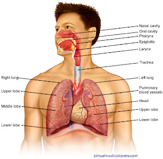
Nose, entrance of external respiration

Nose, entrance of external respiration
Nose is a structure made of bone, cartilage, muscle and epithelium covering. Its skin is consists of numerous sebum gland and hair follicles. Its skin in front, cross opening of the nose and extend to vestibule of the nose.
Here its lining epithelium is condensed and squamous and has strong hair follicle.
Internal cavity is known nasal cavity which is separated into right and left side by a thin wall. Lining epithelium of the cavity is siliated, contains mucous membrane and blood vessels and nerve supply. Upper part of the nasal cavity is  lined by mucous membrane that contains nerve cell of Olfactory (Central nervous system).


Path of respiration

Activities of Nose:
1. Help in entrance of external air in respiration.
2. In case of respiration, purification of the amount of air from dust, micro-organism, which is done by nose.
3. During respiration air conditioned and become warm and moisture which is easy to cross the path of  respiration.

Screening of Breast Cancer

Breast cancer is number one cancer problem in world wide. If this cancer is detected in primary stage then the rate of death can be decrease.  In primary stage the cancer can be detected by a method named ‘Breast cancer screening’. By examining breasts searching of sign of cancer when any disorder or sign is not present in breast. Maximum time successful treatment is done by not surgical removal of breast.
screening of breast cancer

For which women screening is needed??

A woman by born is in risk of breast cancer. But all women are not in same risk. Generally there is no risk before 20 year old. The risk of cancer begin being old. When age is near to the 50 then risks is increased. In case of both highly risked and low risked women the screening test should be done by considering some risk factor.
But in case of highly risked women the screening test should be done from early age and every year.
Low risked women
Woman who has no sign and symptoms of cancer.
In whom family background is absent like mother, sisters of mother, grandmother, daughters have not affected by cancer. Or any member of family has no history of cancer like ovary cancer or colon cancer.
*       Body weight is normal, habit of regular exercise or workout.

*      
Breast feeding of baby for 18 months or more.
High risked women
·        If any one of female family member or relative are affected by breast cancer especially history of more than one of relative. Also history of other cancer.
·        Previous record of breast cancer or primary stage of it were detected in past.
·        If there is excess number of mammary gland (Breast is consist of mammary gland).
·        Excess fat, weight and avoidance of hard working.
·        If period or menstrual cycle occur in early age and Menopause occur in late age.
**
**·        Women who never conceive a baby.
·        Avoidance of breast feeding of baby. 
·        Taking of O.C.P for a long time or Hormone therapy after Menopause.

What are tests done in screening???

Breast Self examination (B.S.C)
It is done by examination of breasts by self and start it from age of 20 year in every month. And continue it for whole lifetime.The aim of this examination is to detect any abnormality of breast and take appropriate treatment for it. Abnormalities like feeling of any mass in breast, thickness of skin of breast, secretion of any liquid or blood from nipples.

self examination of breast 

Clinical Breast examination (CBC)

The method is clinical examination of breasts by trained doctors. One examination is done in 3 years at age of 20-39 year and every year after 40 age of year. Beside this, Doctors advice to do other examinations.
 
Mammography and other radiology and imaging examinations
 Ultrasonography, MRI and if necessary then F.N.A.C (Fine Needle Aspiration) that is aspiration of fluid from affected side by needling and microscopic examination.


mammography
What is Mammography??
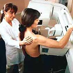
Mammography is an X-ray method of breast examination. Especial type of machine is used in it. In case of using X-ray, radiation is a harmful ingredient of causing breast cancer. But in Mammography a little amount of radiation is used. After 50 years age, in interval of 2-3 years if there done screening, mammography and in primary stage the disease is detected then the benefit is more than harm.So, Mammography is the strongest media. Under specialized radiologist, female radiology technologist operate mammography machine and give the reports. Generally after 50 year mammography should be taken and if health condition is well then continue it for 70 year in 3 year interval. In developed country, different committee held this type of conference for a long time. Some of them advised to do screening mammography for 47 to 73 years old. In case of highly risked women it is suggested after 40. Under 30 it is not applicable because in younger women the mammary glands are so concentrated. So if any mammography is done then the image can be hidden under these

Ladies fast in using emoticons...


Happy
In mobile, sending S.M.S or massages, ladies use emoticons more than men. But in case of using various emoticons men are first. Recently in a survey it is discovered.
Using different letters and sign we use emoticons. These are used to express our different condition and mood. We use it usually in chatting or S.M.S.
Angry
In America, researchers of Rise University of Texas gave a Smart phone in free of cost to every man and woman. But they didn’t tell the people about the research. Then total 10 million and 24 thousand S.M.S are researched. In this case, observed that all of them use emoticons but not in same way. They used it 4% in total sending S.M.S. And ladies are using double emoticons than men. The result of this research has published in 'Computer in Human Behavior Magazine'. ;)